Bursa E-Max veneer applications, medical information about the biological structure of lithium disilicate ceramics, their differences from zirconium, and clinical treatment stages.
Oral and dental health has a direct effect on individuals' physiological integrity and psychological well-being. In cases where a healthy enamel layer has been structurally damaged, shape abnormalities are present, or advanced discoloration has occurred, dentistry aims to repair lost tissues with biocompatible materials. Thanks to advances in dental material engineering, all-ceramic restorations that can precisely mimic the light transmittance and physical resistance of natural teeth are coming to the forefront.
If you have structural loss, gaps (diastema), or aesthetic concerns in your front teeth and are conducting an objective, clinical investigation into Bursa E-max veneer procedures at a medical level, this guide will allow you to examine the biological structure and treatment stages of lithium disilicate ceramics in detail.
This article has been prepared in accordance with Law No. 1219 and healthcare information regulations in order to inform the public about aesthetic and restorative dentistry practices.
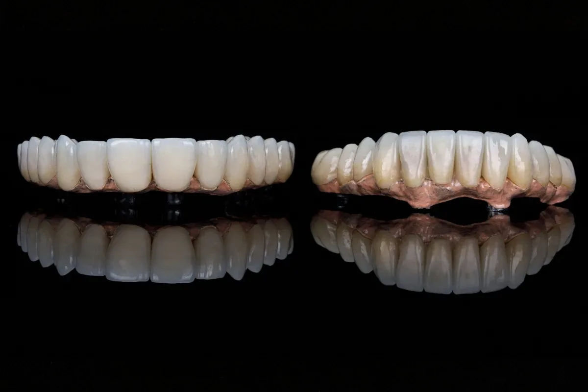
Known in dental terminology as an abbreviation of the words Empress Maximum, E-Max is a reinforced porcelain type containing lithium disilicate glass ceramic and no metal support in its substructure. It is obtained by pressing or milling (carving) naturally occurring crystalline ceramics at very high temperatures in special laboratory furnaces.
Natural tooth enamel does not completely block light; while it reflects some of it, it transmits much of it to the underlying layers (dentin). The main reason E-Max restorations are so widely accepted in the medical world is that their glass ceramic matrix perfectly mimics this translucency feature. Since it does not contain a metal or opaque (matte) zirconium core, it offers not an artificial whiteness but a natural depth that integrates with the tissue when applied.
The material to be used in repairing tooth loss or damage is determined by the dentist's radiographic and clinical examination. The structural differences E-Max ceramics offer compared with other dental materials are as follows:
Light Transmittance (Aesthetic Integrity): Metal-supported porcelains completely block light. Zirconium, on the other hand, offers a durable white substructure, but it is not as translucent as E-Max. In front teeth, where aesthetics are the primary priority (incisors and canines), E-Max provides the highest translucency value.
Mechanical Resistance (Flexural Strength): Zirconium material has higher resistance than E-Max against flexing and chewing pressure. For this reason, zirconium is generally preferred in posterior (molar) teeth or long bridges with multiple missing teeth. E-Max, however, is safely used in single-tooth veneers and aesthetic restorations in the front region.
Tissue-Friendly Structure (Biocompatibility): Lithium disilicate ceramics are compatible with human tissue at the cellular level. They do not cause allergic reactions in the gums, inflammation, or the purplish corrosion line seen in metal restorations. Their plaque retention rate is even lower than that of natural tooth enamel.
The resolution of structural problems in the smile line with bioprotective (least damaging to tooth tissue) methods is the basis of current clinical approaches. Bursa E-max veneer procedures are planned by dentists in line with the following medical needs:
Cases of Wide Diastema (Gapped Teeth): When the spaces between front teeth, whether congenital or acquired later, need to be closed without orthodontic treatment.
Enamel Defects and Fractures: In the repair of teeth whose structure has weakened and aesthetics have been lost due to trauma, teeth grinding, or developmental enamel disorders (hypoplasia).
Resistant Internal Discoloration: In teeth that have lost vitality after root canal treatment and turned gray, or teeth that have become internally discolored due to the use of tetracycline-derived antibiotics during childhood.
Mild Crowding: In situations where the patient is not suitable for orthodontic (braces/clear aligner) treatment or refuses it, for the aesthetic masking of mild alignment irregularities.
Revision of Old Restorations: In replacing old metal-supported crowns or large composite fillings that leak due to incompatible margins and harm gum health.
The production and application of all-ceramic restorations to the patient requires high precision (micron-level) and a disciplined clinical protocol. A standard E-Max treatment consists of the following clinical stages:
Before the procedure, the health of the tooth roots, bone level, and periapical tissues (root apex) are evaluated with panoramic and periapical X-rays. If there is an active infection in the gums (gingivitis or periodontitis), periodontal treatment (gum treatment) is applied first, because precise impressions cannot be taken on a bleeding surface and bonding (cementation) cannot be successful.
The relevant teeth are numbed under local anesthesia. To allow the E-Max restoration to fit the tooth with a natural form, a thin circumferential reduction (preparation) is made from the enamel layer. This reduction is usually between 0.5 mm and 1.5 mm. A clear finish line is created at the gum margin using special bur techniques called stepped reduction. Because the patient is under anesthesia, they feel no pain or soreness.
Once the teeth are brought to the appropriate geometric form, data transfer to the prosthetic laboratory begins. In advanced clinics, this process is converted into a three-dimensional digital impression within seconds using intraoral optical scanners (CAD/CAM intraoral scanner). When necessary, traditional silicone-based impression materials are also used with high precision. Face anatomy and lip dynamics are photographed and sent to the laboratory.
Because the teeth whose enamel layer has been reduced have exposed dentin canals, they are highly sensitive to thermal (hot-cold) stimuli. To prevent this and enable the patient to continue social life without aesthetic concern, acrylic-based temporary teeth are produced and attached to the tooth by the dentist during the same session.
Based on the data taken in the laboratory, the digitally designed E-Max blocks are milled on milling devices or shaped with the press casting technique. During the "substructure and dentin try-in" stage performed in the clinic, the contact points of the veneers with adjacent teeth, the gingival margin (edge fit), and the occlusal (bite) balance during chewing are checked. After color characterization is completed, the restorations are given a glass-smooth surface (glaze).
One of the greatest advantages of lithium disilicate ceramics is that they can bond not only mechanically but also chemically to the tooth at a microscopic level. In this process called "adhesive cementation," the tooth surface and the inner surface of the porcelain are roughened with special acids (hydrofluoric acid, etc.), bonding agents (silane and bond) are applied, and the restoration is sealed to the tooth with special light-cured resin cements. This strong bond eliminates the risk of the crown coming loose.
The outer surface of all-ceramic restorations is so smooth that bacteria cannot adhere to it, and they do not change color from foods and beverages such as tea, coffee, or wine. However, regular care is essential to prevent decay in your underlying natural tooth tissue and to preserve gum health:
Brushing should be done twice a day with the correct technique (sweeping from the gums toward the tooth) and with fluoride toothpaste.
To prevent food buildup and plaque formation at the interfaces of the veneers, interdental brushes or dental floss must absolutely be included in the daily routine. Using dental floss will not dislodge the E-Max; on the contrary, it preserves gum health and extends the life of the restoration.
Chewing habits should be observed; hard-shelled foods such as hazelnuts and walnuts should not be cracked with the teeth. E-Max is a durable ceramic, but uncontrolled, traumatic forces can cause small chips (chipping), as with any porcelain.
If there is a teeth-grinding (bruxism) problem, transparent occlusal splints (night guards) designed by the dentist should be used consistently to prevent excessive load on the ceramic surfaces during sleep.
As with all dental restorations, 6-month dental checkups should become routine.
Lithium disilicate (E-Max) ceramics, which combine aesthetics, biocompatibility, and durability in a single material, become functional tissues that the patient can use safely for life with proper medical planning and precise clinical workmanship. The essential step is to undergo a physical examination at a fully equipped dental clinic to determine the right treatment protocol suited to your own anatomy.
This page content has been prepared solely to medically and objectively inform the public about oral and dental health practices and restorative materials within the framework of the provisions of Law No. 1219 on the Practice of the Art of Medicine and Its Branches and the Ministry of Health's Regulation on Promotional and Informational Activities in Health Services. The information provided herein cannot be used alone for the diagnosis or treatment of diseases, does not contain medical guarantees such as "definite aesthetic success," and does not aim to provide commercial guidance or unfair competition. To scientifically determine the most suitable treatment plan for your oral, dental, and jaw structure, you must consult a dentist directly and undergo an in-person clinical examination.
Let us take a look together and go over your treatment options. Feel free to call us with any questions.
 Türkçe
 English